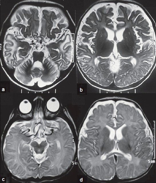
Figure legend from original publication:
Magnetic Resonance images of patients with Menkes disease showing the spectrum of imaging findings:
Axial T2-Weighted images of patient 3 (a and b) show the tortuosity of vessels with
hyperintense signal changes of white matter with tendency to cyst formation at temporal poles
and marked cerebral and cerebellar atrophy.
The images of the patient 5 (c and d) shows marked tortuosity of intracranial vascular flow voids
with mild white matter signal changes and brain atrophy
Jain P et al. (2014) Menkes disease – an important cause of early onset refractory seizures. J Pediatr Neurosci 9(1):11-16. Full text on PubMed.