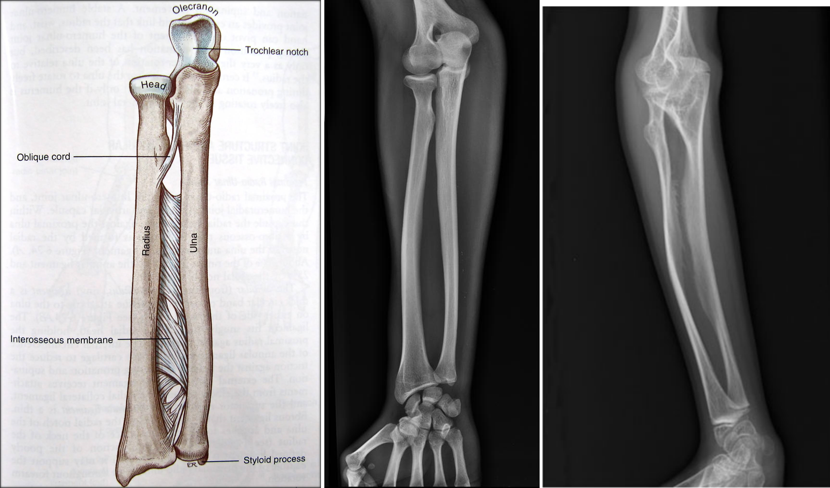

Notes about the figures:
Left. Basic anatomy of the forearm. The forearm has two bones, the radius
and the ulna. The interosseous membrane (IM) is a thin sheet of
Center. An X-ray of a person with a normal
interosseous membrane. Source: Radiopedia (see radiopedia).
Right. An X-ray of an 11-year-old girl with OI type V. The X-ray shows calcification
of her interosseous membrane. Source: below.
Source: Fujino T et al. (2010)
Sporadic osteogenesis imperfecta type V in an 11-year-old Japanese girl.
J Orthop Sci 15(4):589-593. Go back to main osteogenesis imperfecta (type V) syndrome page
tissue that connects them. The IM is important because it creates stability and distributes weight across
the two bones when the arm is being
used. In OI5, the IM becomes calcified, meaning that soft tissue is replaced with calcium. The IM
becomes stiff, and patients have difficulty
rotating their palms up and down. The arm becomes painful and unstable, and the head of the radius (top left
in drawing) may slip out of place.
Figure reprinted with permission from Elsevier.
Abstract on publisher website.
Abstract on PubMed.