Ataxia with Vitamin E Deficiency (AVED)
Ataxia with Vitamin E Deficiency (AVED) is a member of a group of disorders called hereditary ataxias. Hereditary ataxias are are characterized by gait abnormalities. Other problems include hand and speech clumsiness, and abnormal, jerky eye movements. All of these problems derive from abnormalities in the central and/or peripheral nervous systems. Specifically, there are often problems in the cerebellum, the spinal cord, and/or the peripheral nerves. These diseases are progressive, which means that they get worse with time.
There are many hereditary ataxias; FRDA is the most common. Others include ataxia-telangiectasia (A-T), which is the second most common member of the group. Other diseases in the group are categorized in subgroups, such as the spinocerebellar ataxias and the autosomal dominant cerebellar ataxias. Each subgroup has many members. For example, there are so many spinocerebellar ataxias, they are named with numbers (e.g. spinocerebellar ataxia type 1, spinocerebellar ataxia type 17, etc.).Other conditions are named for their clinical features (e.g. cerebellar ataxia, deafness, and narcolepsy, autosomal dominant).
AVED is very rare and estimates for its prevalence vary. A study in Italy found a prevalence of roughly 1 person in 285,000 (1). A study in France found a prevalence of 1 person in 800,000, based on a single case there (2). It has been estimated to occur in Norway with a frequency of 0.6 per million inhabitants (3). It may be more common in North Africa, with a study noting that it was diagnosed in 20% of patients with a Friedreich ataxia-like disease (4). The increased prevalence of AVED in this region may be due to a founder effect and a high rate of consanguinous marriages (e.g. first-cousin marriages).
Like FRDA, AVED has been documented in people of European, Middle Eastern, and North African descent. However, unlike FRDA, AVED has been documented in east Asians, with several reports of affected Japanese individuals (5-8). Consequently, a diagnosis of Friedreich ataxia in a person of Japanese origin may be suspect, and testing for vitamin E deficiency may be wise. Other conditions in the differential diagnosis for both disorders are listed below in the section called Differential Diagnosis.
Clinical information
AVED can be a severe disorder presenting in young childhood to a disorder with symptom onset in the 30s or later. The youngest cases recognized have been infants, and the oldest patient was in his 60s at symptom onset (9). In general, earlier onset is associated with quicker progression of disease and later onset is associated with slower progression. The clinical features of AVED are often very similar to those of Friedreich ataxia, and the two disorders are almost indistinguishable clinically in many cases.
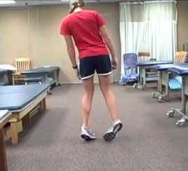
Overall, in milder cases, patients may have slight neurological impairment, while in severe ones, they may resemble FRDA patients (10). Both typically present with progressive ataxia in which patients develop an uncoordinated walk and may experience balance problems. Patients often complain of feeling tired and uncoordinated (10). The most common presenting features of AVED are listed below (data gathered from references 11-24; some patients had more than one presenting symptom):
- Gait ataxia (88/101 patients)
- Titubation (17/101)
- Dysarthria (15/101)
- Clumsiness (12/101)
- Vision impairment (11/101)
Although the age of symptom onset varies overall, there are groups in which it is relatively stable. For example, Tunisian and Moroccan patients typically develop abnormalities around age 13, while North American, European, and Mediterranean patients experience their first symptoms when under age 20 (10).
The most common signs and symptoms of AVED are listed below. As noted, they are very similar to those in Friedreich ataxia. Like FRDA, AVED does not cause a common facial appearance.
- Ataxia (truncal and limb types)
- Absent or reduced tendon reflexes
- Extensor plantar reflexes (Babinski's sign)
- Peripheral neuropathy
- Speech disorder, most notably dysarthria
- Impaired joint position sense (patient does not know the position of a joint when he cannot see it)
- Impaired vibration sense (patient cannot feel a vibrating object)
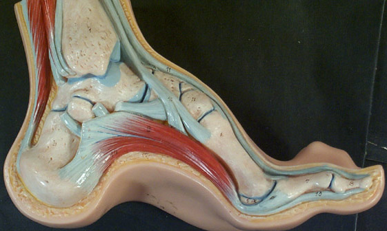
- High arches (pes cavus); may get worse with time
AVED is treated with high doses of vitamin E. The goal of therapy is to normalize serum levels of vitamin E. In most patients, supplementation with 800-1200 mg per day is sufficient (4, 10), but higher doses may be required in some (25). Although prognosis is unpredictable, earlier treatment may lead to better outcomes. For example, in one study of 24 Tunisian patients, intention and head tremor improved over the course of the first year of treatment, and improvements were most obvious in patients who had been affected for less than 15 years (26). No patients in this study deteriorated during treatment. However, the follow-on period was only one year. Alternatively, in a long-term study of 16 Italian AVED patients, 1 improved with treatment, 8 remained neurologically stable, and 7 deteriorated (25). Duration of treatment ranged from 2 to 13 years in this group. The patient whose symptoms had improved was 13 when treatment started and had been symptomatic for only 5 years. Otherwise, there were no correlations between age at onset, timing of treatment onset, length of treatment and outcomes, even among siblings. Similar outcomes have been described in a large cohort of treated patients (14).
Diagnosis and Testing
AVED is an autosomal recessive disorder caused by mutations in the gene tocopherol alpha transfer protein (TTPA). The term autosomal recessive means that the disorder is passed on when both parents contribute a copy of the mutated gene to their child. The link at the right provides information about labs that test for mutations in TTPA.
AVED can be suspected but not diagnosed based on clinical features. This is because of its close similarity to Friedreich ataxia and abetalipoproteinemia (ABL;see below). It is distinguised from FRDA by low serum levels of vitamin E and from ABL by abnormalities in the lipid profile and deficiencies of other vitamins (see Differential Diagnosis). Testing for genetic mutations after laboratory workups can help make a definitive diagnosis.
Unlike FRDA, there are currently no defined signs and symptoms that define AVED. In general, the disease is suspected in a patient who fulfills the criteria for Friedreich ataxia, with the caveat that mild forms of AVED may not mimic FRDA. For the purposes of information, the diagnostic criteria for FRDA are listed below (27, 28). For typical cases of FRDA, diagnosis requires that the following occur within 5 years of symptom onset:
- The patient is less than age 25 at symptom onset (this criterion does NOT apply to AVED)
- Progressive ataxia (limbs and gait) is present
- Knee and ankle jerks are absent or reduced
- Plantar responses are extensor (Babinski's sign)
- Motor nerve conduction velocity is >40 m/s, with reduced or absent sensory nerve action potential
Differential Diagnosis
There are several conditions in the differential diagnosis for AVED. The most important of them are Friedreich ataxia and abetalipoproteinemia. FRDA is not treatable, while abetalipoproteinemia is. The treatment for abetalipoproteinemia requires a low fat diet and supplementation with vitamins A, E, and K.
Friedreich ataxia (FRDA) As noted, AVED and FRDA share many similarities and can be clinically indistinguishable in some cases. However, several authors have identified some differences between them. The key distinguishing feature is that levels of serum vitamin E are normal in FRDA and low or very low in AVED. In addition, diabetes or abnormalities in blood sugar occur in roughly a quarter to a third of FRDA patients. This problem is rare in AVED (10); the same is true of cardiomyopathy. In our literature survey, we found that cardiomyopathy was reported in 57% of ~300 patients whose case histories we analyzed. This problem occured in only 13% of ~100 AVED patients. Thus, although the presence of diabetes and/or cardiomyopathy does not rule out AVED, their presence is more suggestive of FRDA in a patient who is otherwise not at risk for these diseases. Amyotrophy and muscle weakness occur in FRDA but do not appear to affect AVED patients (4).
Alternatively, AVED patients may have head titubation and retinitis pigmentosa. Both of these problems are rare in FRDA. In addition, AVED occurs in east Asians (e.g. people of Japanese origin), while FRDA does not.
Abetalipoproteinemia (ABL). ABL, also called Bassen-Kornzweig syndrome, affects the body's ability to use fats that have been eaten. This is because people with ABL cannot transport fats out of their intestines. Like FRDA, ABL is an autosomal recessive disorder and is very rare. Prevalence is estimated at less than one person per million (29). People with ABL have very low levels of total and LDL cholesterols, and very low levels of triglycerides. These molecules are necessary for the absorption of vitamins A, E, and K from the intestines, and the lab tests show low levels of these vitamins. Vitamin E deficiency causes neurological signs that are similar to those of Friedreich ataxia and AVED (10). However, unliked AVED and FRDA, neurological signs are not the first problems to appear, a fact that can help distinguish ABL from them. The first clinical signs of ABL are generally gastrointestinal, and include steatorrhea (fatty stools), diarrhea with steatorrhea, and liver disease. AVED and FRDA do not generally cause these problems. In addition, laboratory studies of serum cholesterol (total and LDL) are normal in AVED/FRDA but abnormal in ABL. Serum vitamin E may be reduced in both AVED and ABL; levels are normal in FRDA.
Another important feature of ABL that distinguishes it from AVED and FRDA is the presence of star-shaped red blood cells (acanthocytosis). These cells are obvious on a blood smear.
ABL is caused by mutations in the gene microsomal triglyceride transfer protein (MTP), and gene sequencing can provide a definitive diagnosis (see link at right).
Charcot-Marie-Tooth disease (CMT). Charcot-Marie-Tooth neuropathy types 1 and 2 (CMT1 and CMT2) also resemble AVED. Medical problems associated with CMT1 and AVED include sensory loss and slow nerve conduction velocity. Like AVED patients, CMT1 patients may have high arches and they may walk with a foot drop. Symptom onset is usually between ages 5 and 25 years. Some CMT patients are clumsy and lose deep tendon reflexes as children. Dysarthria does not appear to be a feature of CMT1 or CMT2, although it has been reported in a few patients with CMTX, a third form of Charcot-Marie-Tooth disease. this fact can help distinguish AVED from these diseases. Unfortunatley,dysarthria and extensor plantar reflexes may develop over time in AVED, meaning that using them as distinguishing features in a newly presenting patient is likely not an effective approach.
Unfortunately, there are may subtypes of CMT1 and 2, with more than 20 different genes associated with them. However, sequencing for FXN and the CMT genes can rule them in or out. In addition, most types of CMT1 and 2 are autosomal dominant, meaning that the presence of an affected parent is suggestive of CMT over AVED.
Malnutrition. A poor or unbalanced diet may induce AVED-like symptoms. Because vitamin E is stored in fats, a previously healthy person must fail to consume vitamin E over many months in order to symptoms to appear. This problem has been documented in people eating Zen macrobiotic/grain-only diets popularized by George Ohsawa. Grain-only diets can lead to nutritional deficiencies and can be hazardous to health.
Certain chronic diseases can also cause manutrition and vitamin deficiencies. They include cholestatic liver disease, short bowel syndrome, cystic fibrosis, and Crohn's disease (4). Patients with vitamin E deficiency may need to change their dietary habits or may need vitamin supplementation.
References
- 1. Zortea M et al. (2004) Prevalence of inherited ataxias in the province of Padua, Italy. Neuroepidemiology 23(6):275-280. Abstract on PubMed.
- 2. Anheim M et al. (2000) Epidemiological, clinical, paraclinical and molecular study of a cohort of 102 patients affected with autosomal recessive progressive cerebellar ataxia from Alsace, Eastern France: implications for clinical management. Neurogenetics 11:1-12. Abstract on PubMed.
- 3. Elkamil A et al. (2015) Ataxia with Vitamin E Deficiency in Norway J Mov Disord 8(1):33-36. Full text on PubMed.
- 4. Schuelke M (2005) Ataxia with vitamin E deficiency. Updated October 13, 2016. GeneReviews [Internet] Pagon RA et al., editors. Seattle (WA): University of Washington, Seattle; 1993-2021. Full text.
- 5. Gotoda T et al. (1995) Adult-onset spinocerebellar dysfunction caused by a mutation in the gene for the alpha-tocopherol-transfer protein. N Engl J Med 333(20):1313-1318 Full text from publisher.
- 6. Iwasa K et al. (2014) Retinitis pigmentosa and macular degeneration in a patient with ataxia with isolated vitamin E deficiency with a novel c.717 del C mutation in the TTPA gene. J Neurol Sci 345(1-2):228-230. Abstract pn PubMed.
- 7. Shimohata AE (1997) Ataxia with isolated vitamin E deficiency and retinitis pigmentosa. Ann Neurol 143(2):273 Abstract on PubMed.
- 8. Yokota T (1997) Friedreich-like ataxia with retinitis pigmentosa caused by the His101Gln mutation of the alpha-tocopherol transfer protein gene. Ann Neurol 41(6):826-832. Abstract on PubMed.
- 9. Yokota T (1987) Adult-onset spinocerebellar syndrome with idiopathic vitamin E deficiency. Ann Neurol 22(1):84-87. Abstract on PubMed.
- 10. Hentati F et al. (2012) Ataxia with vitamin E deficiency and abetalipoproteinemia. Handbook of Clinical Neurology 103:295-305. Abstract on PubMed.
- 11. Angelini L et al. (2002) Myoclonic dystonia as unique presentation of isolated vitamin E deficiency in a young patient. Mov Disord 17(3):612-614. Abstract on PubMed.
- 12. Benomar A et al. (2002) Clinical comparison between AVED patients with 744 del A mutation and Friedreich ataxia with GAA expansion in 15 Moroccan families. J Neurol Sci 162(1):97-101. Abstract on PubMed.
- 13. Benomar A et al. (1999) Vitamin E deficiency ataxia associated with adenoma. J Neurol Sci 198(1-2):25-29. Abstract on PubMed.
- 14. El Euch-Fayache G et al. (2014) Molecular, clinical and peripheral neuropathy study of Tunisian patients with ataxia with vitamin E deficiency. Brain 137(Pt 2):402-410. Full text from publisher.
- 15. Harding AE et al. (1985) Spinocerebellar degeneration associated with a selective defect of vitamin E absorption. N Engl J Med 313(1):32-35. Abstract on PubMed.
- 16. Jackson CE et al. (1996) Isolated vitamin E deficiency. Muscle Nerve 19(9):1161-1165. Abstract on PubMed.
- 17. Kara B et al. (2008) Ataxia with vitamin E deficiency associated with deafness. Turk J Pediatr 50(5):471-475. Full text from publisher.
- 18. Koht J et al. (2009) Ataxia with vitamin E deficiency in southeast Norway, case report. Acta Neurol Scand Suppl 120(Suppl 189):42-45. Abstract on PubMed. Full text on ResearchGate.
- 19. Krendel DA et al. (1987) Isolated deficiency of vitamin E with progressive neurologic deterioration. Neurology 37(3):538-540. Abstract on PubMed.
- 20. Müller KI et al. (2011) Epilepsy in a patient with ataxia caused by vitamin E deficiency. BMJ Case Rep 2011:bcr0120113728. doi: 10.1136/bcr.01.2011.3728. Full text on PubMed.
- 21. Puri V et al. (2011) Isolated vitamin E deficiency with demyelinating neuropathy. Muscle Nerve 32(2):230-235. Abstract on PubMed.
- 22. Roubertie A et al. (2003) Ataxia with vitamin E deficiency and severe dystonia: report of a case. Brain Dev 25(6):442-445. Abstract on PubMed.
- 23. Stumpf DA et al. (1987) Friedreich's disease: V. Variant form with vitamin E deficiency and normal fat absorption. Neurology 37(1):68-74. Abstract on PubMed.
- 24. Yokota T et al. (1996) Retinitis pigmentosa and ataxia caused by a mutation in the gene for the alpha-tocopherol-transfer protein. N Engl J Med 335(23):1770-1771. Full text from publisher.
- 25. Mariotti C et al. (2004) Ataxia with isolated vitamin E deficiency: neurological phenotype, clinical follow-up and novel mutations in TTPA gene in Italian families. Neurol Sci 25(3):130-137. Abstract on PubMed.
- 26. Gabsi S et al. (2001) Effect of vitamin E supplementation in patients with ataxia with vitamin E deficiency. Eur J Neurol 8(5):477-481. Abstract on PubMed.
- 27. Harding AE (1981) Friedreich's ataxia: a clinical and genetic study of 90 families with an analysis of early diagnostic criteria and intrafamilial clustering of clinical features. Brain 104:589-620. Abstract on PubMed.
- 28. Pandolfo M (2012) Friedreich ataxia. Handbook of Clinical Neurology 103:275-294. First page viewable from publisher.
- 29. Kane JP (2001) Disorders of the biogenesis and secretion of lipoproteins containing the B apolipoproteins. The Metabolic and Molecular Bases of Inherited Disease, 8th edition Scriver CR et al., eds. New York, NY, USA, McGraw-Hill; 2717-2752.


