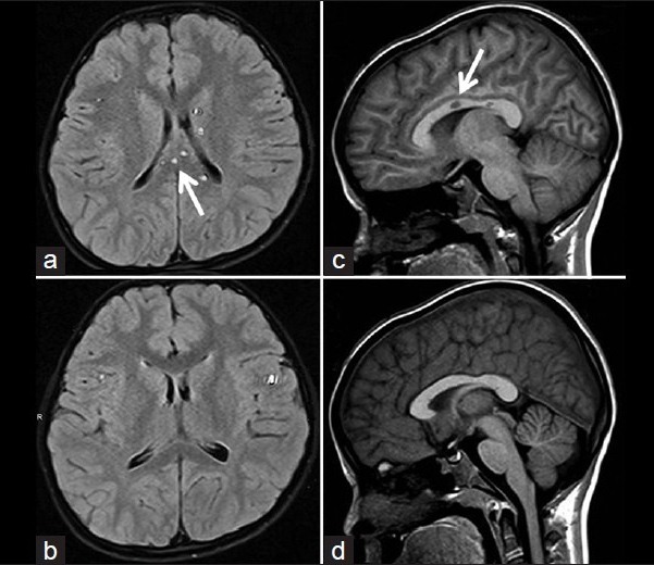
MRI showing typical Findings in Susac's syndrome. Figure legend from original publication:
(a-d) Magnetic resonance imaging FLAIR T2 image showing multiple well-defined hyperintense lesions in periventricular and callosal area.
Magnetic resonance imaging T1 image showing hypointense lesions in center of corpus callosum. Magnetic resonance imaging, 4 weeks later
showing almost complete disappearance of lesions.
Source: Prakash G et al. (2013).
Susac's syndrome: First from India and youngest in the world. Ind J Ophth 61(12):772-773.
Full text on PubMed.
This figure is licensed under the Creative Commons Attribution-Noncommercial-Share Alike 3.0 Unported license, which
permits unrestricted use, distribution, and reproduction in any medium, provided the original work is properly cited.
Back to Susac's syndrome page.