Apert syndrome
Apert syndrome is member of a group of disorders involving craniosynostosis. This term means that at least one of a person's skull bones fuses prematurely. The problem is often noted at birth, but it may be picked up on an ultrasound or it may not become evident until well after birth. Premature fusion of skull bones restricts skull growth, causing it to expand unevenly to accomodate brain growth.
When only one brain suture is closed prematurely, a person is said to have isolated craniosynostosis. This condition is relatively common, occuring in roughly 3-5 per 10,000 births or even more frequently (1, 2). Severity varies widely, with some cases being severe and others being barely noticeable. The causes for isolated craniosynostosis are not fully understood, but hereditary/genetic components are involved (1, 3). Possible risk factors for craniosynostosis include, but are not limited to, the mother's age and race more common among older women and whites); 4), male sex (4), and heavy maternal smoking, especially after the first trimester (5).
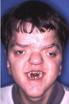
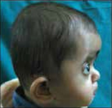
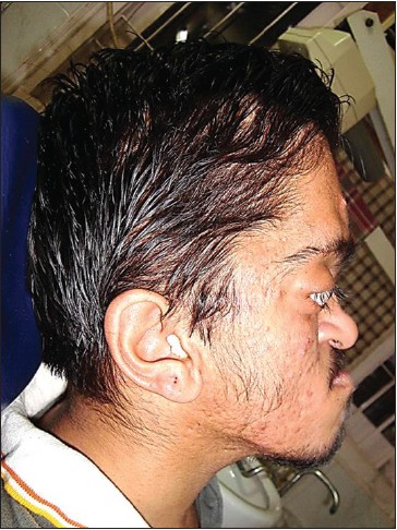
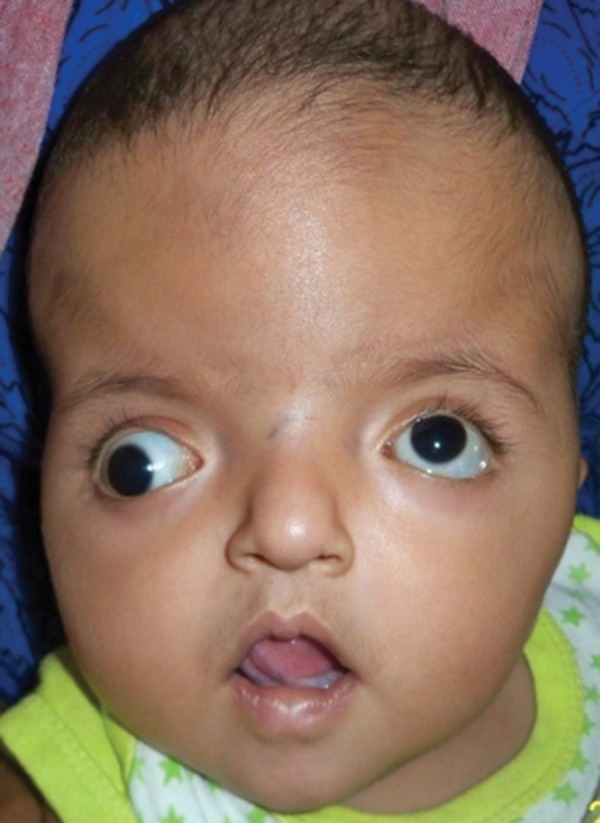
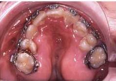
In addition to isolated craniosynostosis, there are also a number of craniosynostosis syndromes. As the name implies, the syndromic forms of craniosynostosis involve problems in a variety of body systems. The hands and/or feet are often affected, a fact that implies that skull and hand/foot development share some type of common mechanism. Apert syndrome is a member of a group named FGFR-related craniosynostosis syndromes. All members of this group are caused by mutations in the genes FGFR1, FGFR2, or FGFR3. In addition to Apert syndrome, the group includes Pfeiffer syndrome, Crouzon syndrome (CS), Crouzon syndrome with acanthosis nigricans, Muenke syndrome, Jackson-Weiss syndrome, Beare-Stevenson syndrome, and FGFR2-related isolated coronal synostosis. Generally speaking, skull abnormalities in these syndromes are observed in the newborn period or before, although some cases may not be noted until later infancy or afterward. In addition to having abnormally shaped skulls, people with these syndromes tend to have wideset eyes that protrude outward (proptosis), an underdeveloped midface (hypoplasia; best seen in profile; see photos on this page), a small nose that may be beaked, and a highly arched palate or a cleft palate (see photos on this page). Intellectual disability is common but does not always occur.
Apert syndrome affects males and females equally. It has also been seen in all races and ethnic groups around the world. In our analysis of ~750 published cases of AS, exactly half were male and half were female. We found reports describing people of European descent, as well as descriptions of Asians (both eastern and western Asia), Latinos, sub-Saharan Africans and African-Americans, Arabs, and others.
Estimates about the prevalence of AS vary from 1 in 42,000 live births in France (6) to 1 birth in 100,000 in a large area of Germany (7) to 1 in 160,000 live births in the UK (8). A study in four countries including four US states found a birth prevalance of 15.5 per million births, or 1 per almost 65,000 (9). This study, given its much larger scale, may have made the most accurate estimate. Regardless, although AS is a very rare disease, it accounts for roughly 4-5% of cases craniosynostosis (9). Crouzon syndrome (see Differential Diagnosis, below) is the most common member of the FGFR-related craniosynostosis syndromes.
Apert syndrome is also called acrocephalosyndactyly type I.
Clinical Information
The cardinal features of AS are craniosynostosis and severe syndactyly of both hands and both feet. These abnormalities are universal or nearly so in AS. Midface hypoplasia is also present in all or nearly all AS patients. This term means that a portion of the middle part of the face is underdeveloped. When viewed in profile, people with midface hypoplasia often appear to have a convex facial structure (see photos on this page). Dental abnormalities are also very common, as is a highly arched or cleft palate; the gums in the area of the palate are typicaly swollen, as well (see photo on this page).
As noted above, most people with AS have an intellectual disability. However, this problem is by no means an absolute in AS, and normal intelligence should not be used to excluse a diagnosis of AS. Intellectual disabilities in AS are generally in the mild to moderate range.
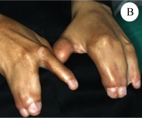
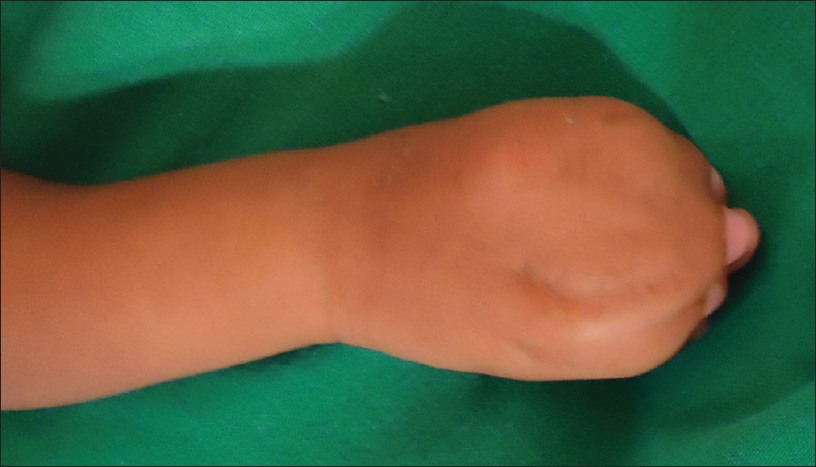
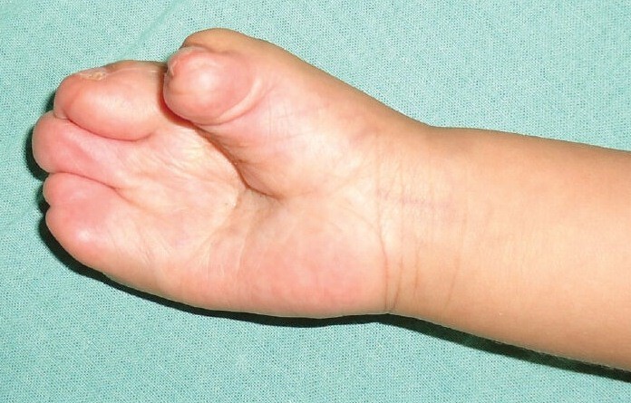
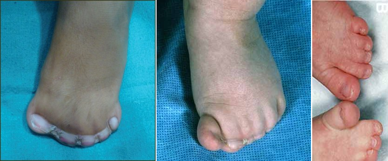
Severe syndactyly of the hands and feet is a consistent and cardinal feature of Apert syndrome. In our literature survey, it was present in all 453 patients for whom information was provided. It occurs in both the left and right hands and feet, and generally involves at least three digits. A classification system has been developed (10, 11); note however, that these categories are not absolute.
- Type 1 syndactyly is the mildest form of this abnormality in AS. In the hand, the central fingers are joined, with the thumb free and the pinky finger partially separated. The thumb is short, wide, and radially deviated. The same pattern can occur in the foot (see photo below).
- In type 2 syndactyly, the thumb is separate, but the other digits are fused. As in type 1 syndactyly, the thumb is short, wide, and radially deviated. The nails may also be fused or partially fused, and the palms may be described as spoon-shaped. Again, the same basic arrangement can occur in the foot.
- In type 3 syndactyly, all digits are joined, and the finger and toenails may also be joined. The palms are more deeply concave than in type 2 syndactyly. There are photographs of type 3 syndactyly of the hands and foot at the bottom of this page.
The following are common clinical features of Apert syndrome. Because AS overlaps with other craniosynostosis syndromes, laboratory testing may be required to make a definitive diagnosis.
- Craniosynostosis (premature fusion of the skull bones, accompanied by unusual skull shape
- Shoulder girdle and elbow abnormalities, usually with reduced range of motion
- Dental abnormalities (crowding, abnormally positioned teeth, etc.)
- Swelling of the tissue of the palate (see photos)
- Highly arched palate (see photos below)
- Enlargement of the brain ventricles
- Syndactyly of the hands and feet
- Acne (may be severe)
- Intellectual disability
- Excessive sweating
- Hearing loss
Common clinical features of Apert syndrome
In addition, people with Apert syndrome often share a similar facial appearance and may have the following facial features; note that many of these features are shared with other members of the FGFR-related craniosynostosis syndromes, including Crouzon syndrome and Pfeiffer syndrome:
- Midface hypoplasia (the cheekbones, upper jaw, and eye sockets are underdeveloped)
- Downslanting palpebral fissures (see photo at top right)
- Hypertelorism/wideset eyes
- Proptosis/protruding eyes
- Facial assymetry
- Large forehead
- Low-set ears
Proptosis can be severe and may impair vision, as can optic nerve atrophy, abnormalities of extraocular muscles, cataracts, ectopia lentis (displaced lens), and other problems (reviewed in 12).
Dental abnormalities are also a very common problem in AS.
Diagnosis and Testing
AS should generally be suspected in a person presenting with craniosynostosis, severe syndactyly as described here, and midface hypoplasia. These three abnormalities occur universally or nearly so in people with AS. Other highly common clinical features of AS include dental abnormalities (crowded teeth, malocclusion and bite abnormalities, late development of teeth, etc.), a highly arched or cleft palate, and swelling of the gum tissue in the area of the palate (see photos on this page).
AS is caused by mutations in the gene FGFR2. It is an autosomal dominant disorder. This term means that only one copy of a mutated gene is needed to cause the disorder. Researchers estimate that 95% of cases of AS are sporadic, meaning that they arise when a mutation in FGFR2 occurs randomly and for reasons that are not known. However, advanced paternal age has been found to be a risk factor for AS (8, 13) and other members of this syndrome group. Overall, however, the sporadic nature of AS means that the risk of having an affected sibling is low (note that there is a 50% chance that an affected adult will pass the syndrome to his or her offspring).
Mutations in FGFR2 also cause Crouzon syndrome, a condition that is similar to AS. The individual mutation appears to drive disease phenotype. For example, two mutations have been found in more than 98% of cases of AS: they are Ser252Trp and Pro253Arg in exon 3a of FGFR2. Neither of these mutations is associated with Crouzon syndrome or the other conditions in this syndrome group. Crouzon results from a variety of mutations, and in some cases, the same mutations cause Crouzon syndrome, Pfeiffer syndrome, Jackson-Weiss syndrome, and/or Crouzon syndrome with acanthosis nigricans (14). Researchers do not currenly understand why the same mutations lead to different syndromes.
Because the vast majority of AS cases are associated with two mutations that are not associated with any other disorder, genetic sequencing is highly sensitive for Apert syndrome. Clicking the link at the upper right part of this page will link to information about laboratories and other facilities that perform testing. There is also a link for generalized testing of craniosynostosis. As noted in the last paragraph, however, the same mutations may cause different syndromes, meaning that the sensitivity of molecular testing is lower for other craniosynostosis syndromes. The best use of sequencing may be to to help confirm or reject a questionable diagnosis that is based on clinical features only.
Differential Diagnosis
The differential diagnosis for AS includes certain other craniosynostosis syndromes.
Crouzon syndrome (CS). Crouzon syndrome is the most common of the craniosynostosis syndromes, and is also very similar to AS. People with both syndromes have craniosynostosis, wideset eyes, proptosis/protruding eyes, and dental abnormalities. Unlike Apert syndrome, however, Crouzon syndrome does not generally affect the hands and feet. Given that syndacytly is universal in AS, this clinical feature can help distinguish the two conditions. In addition, most patients with Crouzon syndrome do not have intellectual disabilities (11% with IQs <80 in our analysis), while a majority of AS patients do (~75% in our analysis).
Pfeiffer syndrome (PS). Pfeiffer syndrome is another craniosynostosis syndrome. It is also similar to AS. People with PS have craniosynostosis, foot syndactyly, proptosis/protruding eyes, highly arched palates and dental abnormalities. They may also have facial asymmetry and downslanting palpebral fissures. Intellectual disability occurs in PS, but intelligence is normal in the majority of patients. Unlike Apert syndrome, however, PS does not generally cause syndactyly of the hands. Given that finger syndacytly is universal in AS, this clinical feature can help distinguish the two conditions.
Jackson-Weiss syndrome (JWS). JWS is a very rare member of FGFR-related craniosynostosis syndromes group. It was reported in a large Amish kindred in 1976 and is also similar to AS (15). As with Crouzon syndrome, people with JWS and AS have craniosynostosis, wideset eyes, proptosis, and foot abnormalities. Unlike Apert syndrome, however, JWS does not generally cause syndacytly of the hands. Given that this abnormality is universal in AS, its absence can help distinguish the two conditions. Relatively few studies have reported on JWS; the largest did not find evidence of intellectual disability, although it has been reported in a smaller study (16).
Saethre-Chotzen syndrome (SCS). Although SCS is a syndrome involving craniosynostosis, it is not a member of the FGFR-related craniosynostosis syndromes group, because it is caused by mutations in the gene TWIST1. SCS is an autosomal dominant disorder, but unlike in AS, most people with SCS have an affected parent (17). The clinical features of SCS vary, even in affected members in the same family. Severity ranges from mild to severe. Most people who have SCS have craniosynostosis, although the cranial cutures that are fused vary. Strabismus (misaligned eyes) is also common. Syndactyly may be present, but it is not as severe as it is in AS and usually affects only two fingers. In addition, the cosmetic features of SCS are usually mild in comparison to AS and most SCS patients have normal intelligence. Severe intellectual disability appears to be rare in SCS (17) and AS.
References
- 1. Cohen MM & MacLean RE, eds. (2000) Craniosynostosis: Diagnosis, evaluation, and management. New York: Oxford University Press.
- 2. Reefhuis J et al. (2003) Fertility treatments and craniosynostosis: California, Georgia, and Iowa, 1993-1997. Pediatrics 111(5 Pt 2):1163-1166. Full text from publisher.
- 3. Boyadjiev SA et al. (2007) Genetic analysis of non-syndromic craniosynostosis. Orthod Craniofac Res 10(3):129-137. Abstract on PubMed.
- 4. Alderman BW et al. (1988) An epidemiologic study of craniosynostosis: risk indicators for the occurrence of craniosynostosis in Colorado. Am J Epidemiol 128:431-433. Abstract on PubMed.
- 5. Carmichael SL et al. (2008) Craniosynostosis and maternal smoking. Birth Defects Res A Clin Mol Teratol 82(2):78-85. Abstract on PubMed.
- 6. Renier D et al. (1996) Prognosis for mental function in Apert's syndrome. J Neurosurg 85(1):66-72. Abstract on PubMed. Full text on ResearchGate.
- 7. Tünte W & Lenz W (1967) Zur häufigkeit und Mutationsrate des Apert-Syndroms (The frequency and mutation rate of Apert syndrome) Humangenetik 4(2):104-111. Abstract on PubMed. First two pages on publisher website
- 8. Blank CE (1960) Apert's syndrome (a type of aerocephalosyndactyly): observations on a British series of thirty-nine cases. Ann Hum Genet 24:151-164. Abstract on PubMed.
- 9. Cohen MM Jr et al. (1992) Birth prevalence study of the Apert syndrome. Am J Med Genet 42(5):655-659. Abstract on PubMed.
- 10. Upton J (1991) Apert syndrome. Classification and pathologic anatomy of limb anomalies. Clin Plast Surg 18:321-325. Abstract on PubMed.
- 11. Cohen MM Jr & Kreiborg S (1995) Hands and feet in the Apert syndrome. Am J Med Genet 57(1):82-96. Abstract on PubMed.
- 12. Khong JJ et al. (2006) Ophthalmic findings in Apert syndrome prior to craniofacial surgery. Am J Ophthalmol 142(2):328-330. Abstract on PubMed.
- 13. Erickson JD & Cohen MM Jr (1974) A study of parental age effects on the occurrence of fresh mutations for the Apert syndrome. Ann Hum Genet 38:89-96. Abstract on PubMed.
- 14. Wilkie AO (1997) Craniosynostosis: genes and mechanisms. Hum Mol Genet 6(10):1647-1656. Abstract on PubMed.
- 15. Jackson CE et al. (1976) Craniosynostosis, midfacial hypoplasia and foot abnormalities: an autosomal dominant phenotype in a large Amish kindred. J Pediatr 88(6):963-968. Abstract on PubMed.
- 16. Cross HE et al. (1969) Craniosynostosis in the Amish. J Pediatr 75(6):1037-1044. Abstract on publisher website.
- 17. Gallagher ER et al. (2003) Saethre-Chotzen Syndrome. Updated June 14, 2012. GeneReviews [Internet] Pagon RA et al., editors. Seattle (WA): University of Washington, Seattle; 1993-2021. Full text.
- 18. Hohoff A et al. (2007) The spectrum of Apert syndrome: phenotype, particularities in orthodontic treatment, and characteristics of orthognathic surgery. Head Face Med 3:10; doi: 10.1186/1746-160X-3-10. Full text on PubMed.
- 19. Saberi BN & Shakoorpour A (2011) Apert syndrome: report of a case with emphasis on oral manifestations. J Dent (Tehran) 8(2):90-95. Full text on PubMed.
- 20. Gupta M et al. (2013) Anterior plagiocephaly in an atypical case of Apert syndrome. World J Plast Surg 2(2):115-118. Full text on PubMed.
- 21. Bhatia PV et al. (2010) Apert's syndrome: Report of a rare case. J Oral Maxillofac Pathol 17(2):294-297. Full text on PubMed.
- 22. Khan S et al. (2012) Apert Syndrome: a case report. Int J Clin Pediatr Dent 5(3):203-206. Full text on PubMed.
- 23. Patel K et al. (2013) Anesthesia management in a patient of Apert syndrome. Anesth Essays Res 7(1):133-135. Full text on PubMed.
- 24. Kumar N et al. (2014) Anesthetic management of craniosynostosis repair in patient with Apert syndrome. Saudi J Anaesth 8(3):399-401. Full text from publisher.
- 25. National Human Genome Research Institute. Elements of Morphology: Human Malformation Terminology (Online encyclopedia).
